
Fourteen elements, measured the same way every visit, with the arithmetic written out. That is the whole standard for wound care documentation. Miss one element and the same note fails three audiences at once: the payer reviewing a debridement or a skin-substitute claim, the colleague who sees the patient next week, and the auditor who asks whether the wound was actually improving.
This guide is the checklist, written for the clinicians and nurses who chart wounds every day:
- The 14 elements a wound note must carry, with the units and scales for each
- The clock method for length, width, depth, undermining and tunneling, and the two formulas (surface area, percent change) that turn measurements into a progress record
- A wound documentation cheat sheet and a copy-ready template you can paste into any EHR
- Two worked examples: an initial pressure-injury visit and a venous-ulcer follow-up with a debridement
- What Medicare looks for in 2026, including the new skin-substitute rules, and the errors that trigger denials
- How AI dictation changes the work, and what it cannot do
DeepCura publishes this guide and is one of the tools discussed in the last section. It was written from the public standards cited in the references (NPIAP, WOCN, AHRQ, CMS) and has not yet been reviewed by an independent wound-care clinician; treat it as a documentation checklist, not clinical guidance, and follow your own program's protocols and your Medicare Administrative Contractor's coverage policies. No prices are quoted. The note screenshot is a fictional patient rendered in DeepCura's real note view. Where a wound-specific EMR or imaging tool fits a practice better than an AI scribe, this guide says so.
The 14 elements every wound note must carry
Wound care documentation is unusual among specialties: most of the note is structure and numbers rather than narrative. A good wound note reads the same at every visit, in the same order, so that a reader (or a spreadsheet) can find each element in the same place and compare it with the last visit. The table below is the consolidated list from the NPIAP, WOCN and AHRQ guidance in the references, with the unit or scale that makes each element comparable over time.
| # | Element | What to record | Unit or scale |
|---|---|---|---|
| 1 | Location | Anatomic site with laterality (left lateral malleolus, sacrum, right ischial tuberosity) | Body map term |
| 2 | Etiology and type | Pressure, venous, arterial, diabetic/neuropathic, surgical, traumatic, moisture-associated, mixed | Named cause |
| 3 | Classification or stage | Pressure injuries: NPIAP stage 1 to 4, unstageable, deep tissue. Diabetic foot: Wagner or WIfI. Venous: CEAP | System named in the note |
| 4 | Onset and duration | Date first noted, days or weeks present, acquired before or during the admission | Date |
| 5 | Size | Length, width and depth measured the same way each visit | cm, one decimal |
| 6 | Surface area | Length × width, computed and written | cm² |
| 7 | Undermining and tunneling | Depth and clock position of each | cm at o'clock |
| 8 | Wound bed tissue | Granulation, slough, necrotic or eschar, epithelial, hypergranulation | Percentages that total 100 |
| 9 | Exudate | Amount and character (none, scant, moderate, large; serous, serosanguineous, purulent) | Ordered scale |
| 10 | Odor, edges and periwound skin | Odor present or absent; edges attached, rolled, epibole, macerated; periwound intact, erythema, induration, maceration, callus | Descriptors |
| 11 | Signs of infection | Warmth, erythema extent, induration, increased drainage, new pain, fever, or none | Present or absent, with which |
| 12 | Pain | Score and any procedural pain | 0 to 10 |
| 13 | Progress | Condition compared with the prior visit and the baseline: improving, unchanged, stalled, worsening, resolved, plus the percent change in area | Ordered choice and percent |
| 14 | Interventions and plan | Debridement (type, instrument, depth, area, hemostasis), dressings and frequency, offloading, compression, adjuncts, risk score, follow-up interval | Exactly what was done |
Three of these are computed rather than observed: surface area, percent change, and the debridement area that drives the procedure code. Those three are where most documentation breaks down, because they require arithmetic at the bedside or after the visit. The next section gives the rules.
Measuring a wound: the clock method and the two formulas
The clock method fixes the orientation so that measurements are comparable across visits and across clinicians. The patient's head is 12 o'clock and the feet are 6 o'clock, regardless of the wound's location.
- Length is the longest head-to-toe measurement (12 to 6 o'clock), in centimeters to one decimal.
- Width is the widest side-to-side measurement perpendicular to length (3 to 9 o'clock).
- Depth is the deepest point, measured with a sterile cotton-tipped applicator held perpendicular to the wound bed, marked at skin level and read against a ruler.
- Undermining is tissue destruction under intact skin at the wound edge; record the deepest point and its clock position, and the range of positions if it spans several ("undermining 1.5 cm from 1 to 4 o'clock").
- Tunneling is a narrow channel from the wound bed; record its depth and clock position ("tunneling 2.0 cm at 7 o'clock").
Measure with the same method, the same patient position, and the same instrument every visit, and say which method you used. A measurement without a method cannot be compared with the next one.
The two formulas that turn measurements into a progress record:
- Surface area = length × width, in cm². It is an estimate (it treats the wound as a rectangle), but it is the standard comparison figure and payers read it as such. State it as computed.
- Percent change in area = (baseline area − current area) ÷ baseline area × 100. A positive number is a reduction. Report it against the baseline (the first measurement of this episode of care) and against the prior visit.
A worked calculation: a wound measured 4.2 cm × 3.1 cm at baseline (13.02 cm²) and 3.6 cm × 2.5 cm four weeks later (9.00 cm²). Percent change = (13.02 − 9.00) ÷ 13.02 × 100 = 30.9% reduction. Under the 2026 Medicare skin-substitute policies discussed below, the difference between a documented 30.9% and an undocumented "improving" is the difference between a covered application and a denial.
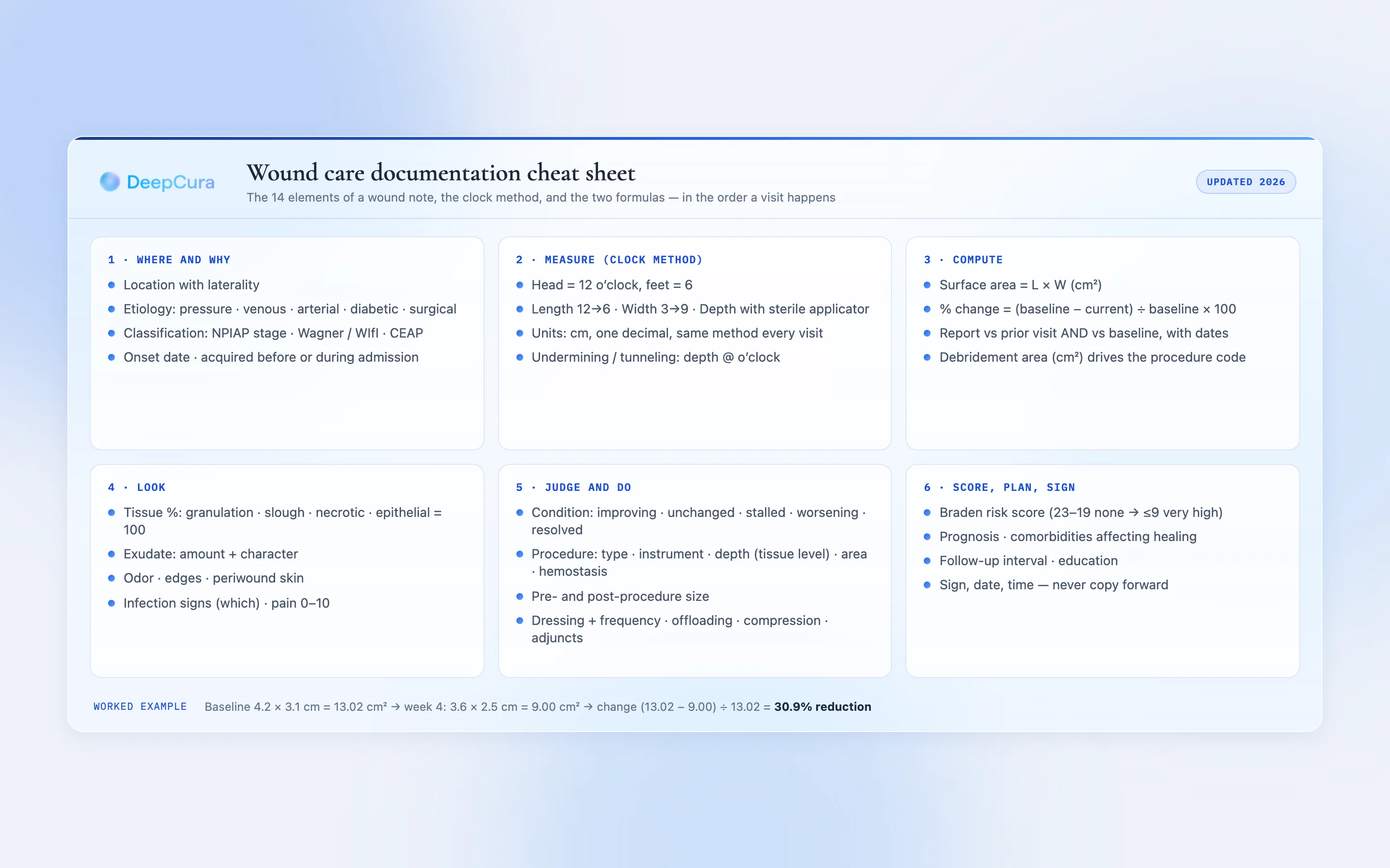
Wound documentation cheat sheet
The same list in the order a visit actually happens, for the exam room:
- Where and why: site with laterality, etiology, classification system and stage, onset.
- Measure: L × W × D in cm to one decimal, clock method stated; undermining and tunneling by depth and o'clock.
- Compute: surface area (L × W) and percent change from baseline and from the prior visit.
- Look: tissue percentages that total 100; exudate amount and character; odor; edges; periwound skin; infection signs; pain 0 to 10.
- Judge: condition versus prior (improving, unchanged, stalled, worsening, resolved) with the number that supports it.
- Do: debridement type, instrument, depth (tissue level reached), area debrided in cm², bleeding controlled; dressing and frequency; offloading or compression; adjuncts.
- Score and plan: risk score (Braden for pressure injury risk), prognosis, comorbidities affecting healing, follow-up interval, patient and caregiver education.
- Sign and date with time; never copy forward a prior visit's measurements.
A wound care documentation template you can copy
This template is written as a fixed block with one label per line, in the same order every visit. Fixed order is not a stylistic choice: it is what allows a spreadsheet, a registry, or a colleague to find each element without reading the whole note. It uses no proprietary wording, so it can be pasted into any EHR's free-text note or used as the instructions for an AI scribe template.
WOUND #1
LOCATION:
ONSET / DURATION:
TYPE / ETIOLOGY:
CLASSIFICATION (system + stage):
WOUND SIZE: Length ___ cm | Width ___ cm | Depth ___ cm | Surface area ___ cm² (L × W)
CHANGE FROM PRIOR VISIT: ___ cm² (___ %) | CHANGE FROM BASELINE: ___ cm² (___ %)
UNDERMINING: ___ cm @ ___ o'clock | TUNNELING: ___ cm @ ___ o'clock
TISSUE: Granulation ___ % | Slough ___ % | Necrotic/eschar ___ % | Epithelial ___ % (total 100)
EXUDATE: amount ___ | character ___
ODOR: ___ | EDGES: ___ | PERIWOUND: ___
INFECTION SIGNS: ___
PAIN: ___ /10
CONDITION: Improving / Unchanged / Stalled / Worsening / Resolved

PROCEDURE (if performed): type ___ | instrument ___ | depth (tissue level) ___ | area debrided ___ cm² | hemostasis ___
POST-PROCEDURE SIZE: Length ___ cm | Width ___ cm | Depth ___ cm
DRESSING / FREQUENCY:
OFFLOADING / COMPRESSION / ADJUNCTS:
RISK SCORE: Braden ___ (23-19 no risk · 18-15 mild · 14-13 moderate · 12-10 high · 9 or less very high)
PROGNOSIS / COMORBIDITIES AFFECTING HEALING:
FOLLOW-UP:
Repeat the block for each wound, numbered in the order they are discussed, and keep the numbering across visits so that Wound #2 this week is the same wound as Wound #2 last week. For a patient with several wounds, add a summary table at the end of the note (one row per wound: current area, prior area, baseline area, percent change, condition). A table is the fastest way for the next reader to see the whole picture.
Related templates on this site: the SOAP note template for the visit narrative around the wound block, and the doctor's note template for work and activity restrictions.
Two worked examples
Both patients are fictional. The examples show the block filled in the way an auditor would want to read it.
Example 1: initial visit, sacral pressure injury
WOUND #1. LOCATION: sacrum, midline. ONSET: first noted 2026-09-12 by facility nursing, present 18 days, acquired prior to admission. TYPE: pressure injury. CLASSIFICATION: NPIAP stage 3. WOUND SIZE: Length 5.6 cm | Width 4.6 cm | Depth 1.2 cm | Surface area 25.76 cm² (L × W). CHANGE FROM PRIOR VISIT: no prior measurement on file; today's area recorded as baseline. UNDERMINING: 1.0 cm @ 2 o'clock; 3.0 cm @ 3 o'clock. TUNNELING: none. TISSUE: Granulation 60% | Slough 40% | Necrotic 0% | Epithelial 0%. EXUDATE: moderate, serosanguineous. ODOR: none. EDGES: attached. PERIWOUND: intact, mild erythema 1 cm. INFECTION SIGNS: none. PAIN: 3/10. CONDITION: new to our service.
PROCEDURE: selective sharp debridement, curette, to subcutaneous tissue, 25.76 cm² debrided, hemostasis with pressure. POST-PROCEDURE SIZE: 5.7 × 4.7 × 1.3 cm. DRESSING: calcium alginate, cover with foam, change every 48 hours. OFFLOADING: low-air-loss mattress, turn every 2 hours, heels floated. RISK SCORE: Braden 14 (moderate). PROGNOSIS: guarded; diabetes and limited mobility affect healing. FOLLOW-UP: weekly.
Example 2: follow-up visit, venous leg ulcer, week 4
WOUND #1. LOCATION: left lower leg, medial gaiter area. ONSET: June 2026, 15 weeks. TYPE: venous insufficiency ulcer. CLASSIFICATION: CEAP C6. WOUND SIZE: Length 3.6 cm | Width 2.5 cm | Depth 0.2 cm | Surface area 9.00 cm² (L × W). CHANGE FROM PRIOR VISIT: −1.98 cm² (−18.0%) from 10.98 cm². CHANGE FROM BASELINE: −4.02 cm² (−30.9%) from 13.02 cm² on 2026-09-02. UNDERMINING: none. TUNNELING: none. TISSUE: Granulation 90% | Slough 10% | Necrotic 0% | Epithelial 0%. EXUDATE: scant, serous. ODOR: none. EDGES: attached, epithelializing at 12 to 3 o'clock. PERIWOUND: hemosiderin staining, no maceration. INFECTION SIGNS: none. PAIN: 2/10. CONDITION: improving.
PROCEDURE: none today. DRESSING: contact layer, absorbent secondary, changed weekly with compression. COMPRESSION: multilayer compression system reapplied; ABI 0.9 on 2026-09-02. FOLLOW-UP: weekly; continue standard care, reassess at week 6 for adjunctive therapy if area reduction stalls.
Notice what the second example does that a narrative note usually does not: it states the prior and baseline areas with their dates, writes the arithmetic, and ties the plan ("reassess at week 6 for adjunctive therapy") to the number.
What Medicare and auditors look for in 2026
Medicare coverage for wound care rests on medical necessity that is demonstrated over time, so the documentation requirements are longitudinal. The elements below are drawn from the CMS Medicare Coverage Database's wound care and debridement local coverage determinations and from the CMS documentation guidance in the references; your Medicare Administrative Contractor's LCD is the controlling text.
- Serial measurements at every visit, with the same method, so that progress toward healing is demonstrable. A note that says "healing well" without numbers is the single most common finding in wound-care reviews.
- Progress toward healing or a documented reason it stalled. When a wound does not improve, the record needs the reassessment: etiology re-examined, adherence, offloading, vascular status, nutrition, infection, and the change in plan.
- Debridement documented as a procedure: the type (selective versus non-selective, sharp versus mechanical or enzymatic), the instrument, the depth as the deepest tissue level reached (skin, subcutaneous tissue, muscle or fascia, bone), the total area debrided in cm², bleeding and how it was controlled, and the pre- and post-debridement measurements. Debridement procedure codes are selected by depth and by total area in 20 cm² increments, so a note without depth and area cannot support the code that was billed.
- Skin substitutes under the 2026 policies. Effective in 2026, Medicare pays for skin-substitute products applied in the office as supplies at a single per-square-centimeter rate, and the contractors' coverage determinations for diabetic foot ulcers and venous leg ulcers require, before the first application, documented standard wound care for at least four weeks with less than a 50% reduction in wound area, plus the measurements that prove both. The determinations also cap the number of applications per episode (eight in the current versions) and require the ongoing measurements that show the product is working. Every one of those conditions is a measurement in the note; none of them can be reconstructed after the fact.
- Medical necessity for the visit itself: the wound's status, the decision-making, and why the service level was needed.
- Legibility, authentication, and no cloned notes. CMS's documentation guidance is explicit that copied-forward entries, identical measurements across visits, and unsigned or undated entries are reasons for denial on review.
- Photographs, when your program uses them, dated, labeled with the wound number, taken with a scale reference, and stored with the patient's consent documented. They support the measurements; they do not replace them.
CPT® codes referenced in this guide are identified by number only. CPT® is a registered trademark of the American Medical Association.
The documentation errors that trigger denials
- A measurement without a method, or a method that changes between visits.
- Length and width recorded without depth, or depth recorded as "shallow."
- No surface area and no percent change, so progress is a word instead of a number.
- Tissue percentages that do not total 100, or "mixed" with no percentages.
- Etiology or classification missing, so the coverage rule that applies cannot be identified.
- A debridement described as "wound cleaned and debrided" with no depth, no area, no instrument and no post-procedure size.
- The same measurements copied forward for three visits.
- A skin-substitute application with no baseline four weeks earlier, or with the standard-care period undocumented.
- "Healing" or "improving" with a larger area than the prior visit.
- Photographs with no date, no wound number, and no consent.
See a wound note compute itself
Dictate the measurements once and watch the surface area, the percent change and the per-wound table appear in the note. Free trial, no credit card.
+1 (415) 549-1829Available 24/7 · Set up in seconds · No credit card required
How AI dictation changes wound documentation, and what it cannot do
Wound care is the specialty where an AI scribe helps least by transcribing and most by computing. The clinician already states the numbers; the burden is the fixed-format block, the arithmetic, the comparison with the last visit and the coding math. That is a different job from the conversation-listening most ambient scribes were built for, and it is why the setup matters more than the model.
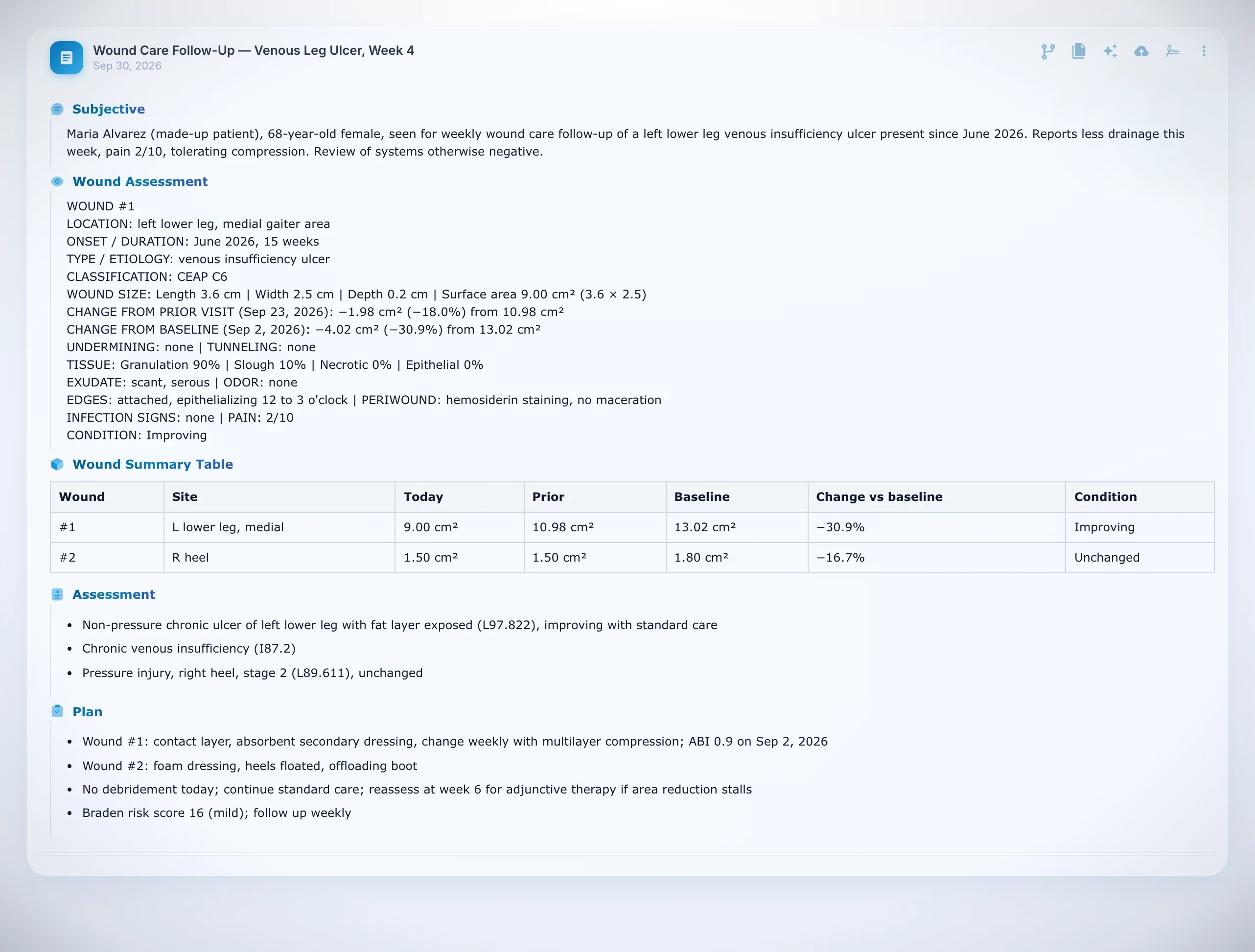
What a well-built wound template does in DeepCura, verified on a live account in September 2026:
- The fixed block, one label per line, in the same order every visit, so a downstream spreadsheet parser or registry finds each element where it expects it.
- Computed lines with the arithmetic shown: the surface area from the dictated length and width, and the change from the prior visit and from baseline in cm² and percent. The prior values come from the patient's tracked measurements, which the clinician confirms in the chart at each visit, or from the previous note attached as context for a same-day or first follow-up.
- A per-wound summary table across visits, rendered as a real table in the note editor.
- Keyword flags: say your own trigger phrase ("debridement macro") and the procedure paragraph is inserted with the post-procedure size and the fields the code needs; nothing is inserted when the phrase is not said.
- Coding arithmetic from the documented depth and area, and site-specific ICD-10 codes with laterality and stage staged for approval, not written silently.
- Per-patient tracked measures (length, width, depth, area, tissue percentages, a risk score) captured from the dictation and confirmed by the clinician, so the trend is on the chart and not in a separate spreadsheet.
What it cannot do, so you can plan around it:
- It does not measure the wound. The clinician measures; the tool records and computes. Imaging apps that measure from a photo are a separate category (see the table below).
- A photo series per wound is not built. One image can be attached per tracked concept per visit; there is no per-wound photo timeline yet.
- The prior-visit arithmetic needs confirmed values. If nobody confirms the tracked measurements in the chart, the next note has nothing to compare against except an attached prior note.
- Rule-heavy templates need a reasoning engine tier, not the fastest one; on the fastest tier the engine tends to drop rules and invent values. The engine choice is a setting the onboarding assistant can change.
- Templates are built per account, from a sample note and the practice's own trigger phrases, on the setup call or with the template tools; there is no pre-built wound library to switch on.
Credit math for a rounding day: at one credit per standard note, a 20-patient skilled-nursing round is 20 credits on the standard engine and 60 on a premium reasoning engine; a facility rounding clinician who records on a phone between rooms and uploads the files afterward pays the same per note and saves the tablet's battery. For the ambient version of the workflow, see ambient dictation and the medical dictation software guide.
Which documentation tool fits which wound practice
No single category is right for every wound program. The table states the fit and the trade-off; where a wound-specific system is the better pick, it says so.
| Situation | Strongest fit | Why | Trade-off |
|---|---|---|---|
| Hospital outpatient wound center with hyperbaric oxygen, registry reporting and program-level analytics | A wound-specific EMR (Net Health WoundExpert, Intellicure) | Built-in wound modules, HBOT documentation, registry exports and benchmark reporting | Another system beside the hospital EHR; per-facility contracts; documentation still typed by the clinician |
| Program that wants automated measurement from photos | Imaging and measurement apps (Swift Medical, eKare, Tissue Analytics) | Calibrated photo measurement and a per-wound image series | The narrative note and coding still live elsewhere; measurement accuracy depends on capture technique |
| Physician practice or rounding group on a general EHR (or copy-paste into a hospital EMR) that dictates measurements | AI documentation with a wound template (DeepCura) | The computed block, the change lines, the table and the coding arithmetic from dictation; tracked measures on the chart; the signed note as a PDF where no integration exists | Does not measure wounds; no per-wound photo timeline; values must be confirmed each visit; not a wound registry |
| Low-volume clinic with a handful of wound patients a month | The EHR's own template plus the cheat sheet above | No new tool to learn | The arithmetic and the comparison are done by hand |
When a practice needs the registry, the hyperbaric module, or photo-based measurement, DeepCura is not the right pick on its own; it is the documentation layer that can sit beside those tools, not a replacement for them. For the four software categories compared by program type, see the wound care EMR and software guide; for the broader comparison of AI scribes across specialties, see the best AI medical scribes guide.
Build your wound template on the setup call
Bring a sample note from your EMR and your trigger phrases; the onboarding assistant builds the template from them. Free trial, no credit card.
+1 (415) 549-1829Available 24/7 · Set up in seconds · No credit card required
Frequently Asked Questions
How do you document a wound?
Record the location with laterality, the etiology and classification, the onset, the size (length, width, depth in centimeters using the clock method), the computed surface area, undermining and tunneling by depth and clock position, tissue percentages that total 100, exudate, odor, edges, periwound skin, signs of infection, pain, the condition compared with the prior visit and baseline, and every intervention with its details. Use the same order and the same method every visit.
How often should a wound be measured and documented?
Measure at every wound care visit and at least weekly for a wound under active treatment, using the same method each time. Serial measurements are what allow progress toward healing to be demonstrated, which is what Medicare coverage for continued treatment and for advanced products depends on.
What is the clock method for wound measurement?
The clock method orients the wound with the patient's head at 12 o'clock and feet at 6 o'clock. Length is the longest measurement from 12 to 6, width is the widest measurement from 3 to 9 perpendicular to length, depth is the deepest point measured with a sterile applicator, and undermining and tunneling are recorded by depth and clock position (for example, 2.0 cm at 7 o'clock).
How do you calculate wound surface area and percent change?
Surface area is length multiplied by width in square centimeters. Percent change is the baseline area minus the current area, divided by the baseline area, multiplied by 100; a positive result is a reduction. Report the change from baseline and from the prior visit, with the dates of both.
What must debridement documentation include?
The type of debridement (selective or non-selective, sharp, mechanical or enzymatic), the instrument used, the deepest tissue level reached (skin, subcutaneous tissue, muscle or fascia, bone), the total area debrided in square centimeters, how bleeding was controlled, the pre- and post-debridement measurements, and the patient's tolerance. Debridement codes are chosen by depth and by area, so a note without both cannot support the code.
What are Medicare's documentation requirements for skin substitutes in 2026?
Under the 2026 Medicare coverage determinations for diabetic foot ulcers and venous leg ulcers, the record must show at least four weeks of documented standard wound care with less than a 50% reduction in wound area before the first application, measurements at each visit, the product and the area applied, and a limited number of applications per episode; Medicare pays office-applied products as supplies at a single per-square-centimeter rate. Confirm the exact criteria in your Medicare Administrative Contractor's local coverage determination.
What is the difference between undermining and tunneling?
Undermining is tissue destruction under intact skin around the edge of the wound, often spanning a range of clock positions; tunneling is a narrow channel that extends from the wound bed in one direction. Both are documented by depth in centimeters and clock position.
Can an AI scribe document wounds?
Yes, if the template is built for it. An AI scribe used for wound care should produce a fixed block in the same order every visit, compute the surface area and the percent change from the dictated measurements and the patient's prior values, insert procedure paragraphs from trigger phrases, and derive coding from the documented depth and area. It does not measure the wound and, in most tools today, it does not maintain a per-wound photo series.
Final Thoughts
Wound care documentation rewards discipline more than eloquence. Fourteen elements, one method, the arithmetic written out, and the same order every visit: that is what lets the next clinician see the trend, what lets a payer approve the next step, and what lets a program prove its outcomes. The cheat sheet and the template above are free to copy into any EHR. If you dictate your measurements and want the computing done for you, the AI-dictation section describes exactly what a wound template can and cannot take off your plate; the best AI medical scribes guide covers the wider field.
References
[1] National Pressure Injury Advisory Panel, "NPIAP Pressure Injury Stages," NPIAP. npiap.com/page/PressureInjuryStages
[2] Agency for Healthcare Research and Quality, "Preventing Pressure Ulcers in Hospitals: A Toolkit for Improving Quality of Care," AHRQ. ahrq.gov
[3] Wound, Ostomy and Continence Nurses Society, clinical practice guidelines and wound assessment resources, WOCN. wocn.org
[4] Centers for Medicare & Medicaid Services, "Medicare Coverage Database," local coverage determinations for wound care, debridement services, and skin substitute grafts/cellular and tissue-based products for diabetic foot ulcers and venous leg ulcers (effective 2026), CMS. cms.gov/medicare-coverage-database
[5] Centers for Medicare & Medicaid Services, "Calendar Year (CY) 2026 Medicare Physician Fee Schedule Final Rule," fact sheet, CMS. cms.gov
[6] Centers for Medicare & Medicaid Services, "Complying With Medical Record Documentation Requirements," MLN Fact Sheet MLN909160, CMS. cms.gov
[7] Lurie F., Passman M., Meisner M., et al., "The 2020 update of the CEAP classification system and reporting standards," Journal of Vascular Surgery: Venous and Lymphatic Disorders, 2020. doi.org/10.1016/j.jvsv.2019.12.075
[8] Mills J.L., Conte M.S., Armstrong D.G., et al., "The Society for Vascular Surgery Lower Extremity Threatened Limb Classification System: risk stratification based on Wound, Ischemia, and foot Infection (WIfI)," Journal of Vascular Surgery, 2014. doi.org/10.1016/j.jvs.2013.08.003
[9] Wagner F.W., "The dysvascular foot: a system for diagnosis and treatment," Foot & Ankle, 1981. doi.org/10.1177/107110078100200202
[10] Braden B., Bergstrom N., "Braden Scale for Predicting Pressure Sore Risk," Prevention Plus. bradenscale.com
[11] American Medical Association, "CPT® (Current Procedural Terminology)," AMA. ama-assn.org/practice-management/cpt